Dental Radiography
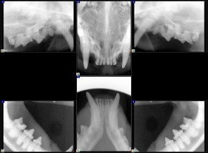
Normal X-Rays of cat’s teeth
Intraoral Radiographs (Dental X-Rays) for Pets
A thorough dental examination includes dental radiographs. More than 50 percent of a dog or cat tooth’s surface area (tooth root) is below the gum line and is therefore not visible to the naked eye. Intraoral radiographs are made using small radiographic films or digital sensors placed inside the patient’s mouth. Since the patient must be very still, intraoral radiographs must be obtained under general anesthesia.
Intraoral radiographs are the best tools to:
- Identify dental disease processes that effect primarily or only the root and its supporting bone. Important pathology can be present that is not apparent from the oral exam.
- Monitor the progress of many previous dental and oral surgical procedures.
Dr. Van Nice is trained in interpreting dental radiographs specifically and is willing to review radiographs on request by primary care veterinarians.
Computed Tomography (CT Scans)
An advanced imaging modality, CT is used to collect images of both hard and soft tissue for viewing in 3 dimensions. This type of imaging is often necessary to determine the extent of certain oral tumors and to determine whether can be completely removed surgically. It is also very helpful in determining the extent of oral fractures and is also a good method for evaluating the temporo-mandibular joint (TMJ).
Magnetic Resonance Imaging (MRI)
MRI is another advanced imaging method that allows both hard and soft tissue to be imaged. It is the preferred method for imaging soft tissues in a dog or cat’s mouth.
CT and MRI are available through our colleagues at Advanced Veterinary Medical Imaging.