Endodontics
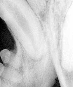
Fractured tooth with necrotic pulp
The “endodontic system” is the hollow area inside a tooth that is filled with sensitive pulp tissue (blood vessels, nerves, and connective tissue), and allows the teeth to grow, mature and respond to stress. The blood supply and nerves enter the teeth through a collection of small holes in the tips of the roots in dogs and cats.
Endodontic disease refers to damage to the dental pulp, commonly termed pulpitis. Depending on the severity of the damage, pulpitis may or may not be reversible. Reversible pulpitis is normally caused by minor trauma, with the tooth surviving the incident. Irreversible pulpitis is a result of inflammation where the tissues swell, preventing blood from entering the root canal and the result is “death” of the tooth. The most common cause of irreversible pulpitis in veterinary patients is fracture of a tooth, exposing the pulp tissue to bacteria in the oral cavity. When this occurs, the inside of the tooth can eventually fill with infected material that can trickle through the openings in the tip of the root into the jaw. The bacteria will then have a secure hiding place inside the actual canal of the root, and the body’s immune system will be unable to resolve the infection, even with antibiotic treatment.
Fractured teeth are a very common occurrence in dogs and cats, usually resulting from external trauma (e.g. when a tooth is hit by any hard object) or vigorously chewing on hard objects. The teeth most frequently broken are the canine (fang) teeth in the dog and the cat, and the upper fourth premolar teeth (the carnassial teeth – the large upper teeth in the back of the mouth) in dogs.
Anyone who has experienced endodontic pain in his or her own mouth can verify that this can be extremely painful. Due to high pain threshold tendencies and other instinctive behaviors, our animal patients rarely show obvious signs of discomfort and will often go to great lengths to hide their pain. The absence of obvious signs of pain can cause an owner to be unaware of or ignore the problem. However, when identified, problems such as a broken tooth or swelling around a tooth should never be ignored, and prompt attention is a must – another reason to have regular wellness exams.
Treating Pulpitis with Root Canal Therapy
A radiograph is an essential first step to evaluate the bone and confirm that the root is intact. There are two options for dealing with a fractured tooth that has exposed the pulp chamber. One option is root canal therapy. This involves removal of the diseased pulpal tissue. The clean and disinfected root canal is then filled with an inert material to prevent future bacterial contamination. Tooth-colored restorations are then placed to seal the crown against further infection. Results of root canal treatment are excellent when the procedure is performed well and with the right equipment. Following completion of a root canal procedure, a radiograph must always be taken to confirm that the canal has been completely filled.
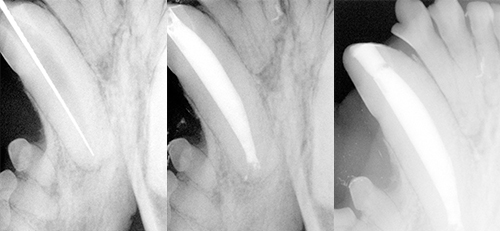
A) Root canal file during procedure
B) Root canal being filled
C) Root canal procedure completed
Vital Pulp Therapy
A related procedure used in immature teeth is vital pulp therapy. This may be performed on recently fractured teeth in younger patients (under 18 months of age). This treatment can help keep the tooth alive, allowing it to become stronger subsequently by laying down new dentin internally. Most veterinary dentists rarely perform this procedure in older patients due to the higher risk of failure when compared to root canal therapy. Teeth treated with vital pulp therapy may require root canal treatment if the vital pulp therapy fails.
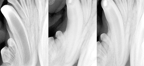
A) Immature tooth being treated by vital pulpotomy
B) Vital pulpotomy has allowed the tooth to mature normally
C) Vital pulpotomy recheck
The only other option for treatment is extraction of the diseased tooth. For the large canine teeth in dogs and cats, and the large chewing (carnassial) teeth in dogs, the extraction procedure can be traumatic and painful due to the size of the roots in our animal patients. The root of the canine tooth is longer and wider than the crown (the part of the tooth above the gumline). Extraction of these teeth involves major oral surgery, comparable to removing impacted wisdom teeth in human patients. The patient also loses the function of the tooth, which can be very important in working dogs. Most veterinary dentists try to avoid extraction of a fractured but otherwise healthy tooth. A metal crown may be indicated following root canal treatment, depending on the extent of crown that is missing and the function of the dog.